At birth, the volume of the brain corresponds to about 30% of that of the adult and reaches 70% at one year of age (L. Konkel, 2018). Very early on, the premises of organisation, already observed in utero, will intensify and be refined through sensory information. Cognitive abilities are gradually established, as the brain develops massively. The first three years are a time when offering a variety of sensory stimulation can be beneficial. By doing this, we can help children to develop their skills without over-stimulation. This is particularly interesting for senses that are not often valued in educational practice: smell, taste, or touch for example. On the other hand, it is useful to help children understand that natural objects are always multi-sensory objects whose different facets must be grasped in order to have a better mental representation. Well-conducted approaches by caregivers can be relevant (in taking into account -and this is important- the age and receptivity of each child, his or her feeling of confidence, his or her ability to focus attention, his or her curiosity, the playfulness of the situations, etc.).
The brain of the newborn is, as in the adult, divided into lobes by deep sulci. In the adult, each lobe is subdivided into topographically identifiable cortical regions whose function is (relatively) known. Schematically, a distinction is made between areas that receive information from sensory sensors (such as the primary visual cortex in the occipital cortex at the back of the brain, or the primary auditory cortex in the temporal lobe) and so-called integration areas, particularly in the parietal lobe, which bring together information from several sensory channels and allow for hypothesised processing. There are also regions that carry out more elaborate tasks such as, for example, the selection of sensory information by directing attention, the pooling of integration areas, the appeal to memorised information, the control of predictions based on available information (with readjustment in the event of error). Others involve emotional feelings. All of these levels of functioning are potentially present at birth, or shortly thereafter, but will only really become established, refined and reinforced through sensory experiences.

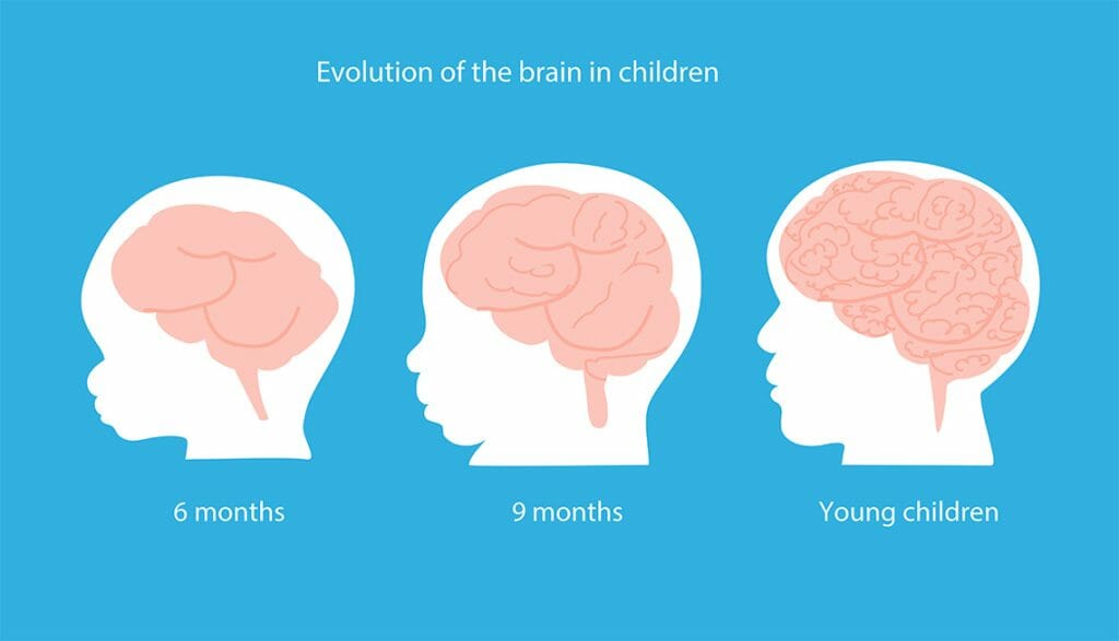
Brain growth is prodigious during the first three years (L. Konkel, 2018). On the one hand, this growth reflects the growth of neurons in the cortex (in the grey matter), the multiplication of synaptic contacts (which allow neurons to exchange information) and the creation of a very large number of communication networks between the different brain regions. On the other hand, the axons of the neurons are surrounded by an insulating sheath (myelin) which increases the speed of transfer of nervous information within the networks. This process of myelination is gradual and takes several years (Sanchez et al (2012). Myelination progresses gradually from the back of the brain to the frontal lobes. The frontal lobes are primarily involved in planning, reasoning, decision-making, language etc., all of which are slowly optimised in the young child.
Different variants of MRI (magnetic resonance imaging) can provide precise information, both structural and functional. Their use to examine the brain at the end of gestation and during early childhood has required great technical ingenuity, because at this period it does not yet have the characteristics of an adult brain. For example, the cortex is still poorly organised, the white matter is poorly myelinated, and various factors complicate the recordings (uncontrollable head movements, higher heart and breathing rates). Very recently, these problems have been overcome (e.g. Cusack et al. (2018)) and we are beginning to better understand how the brain is structured and how the communication pathways between the different cortical regions are set up and allow the exchange of information. Major projects are underway, such as the Baby Connectome Project, to better describe, on a large scale, the evolution of the brain from birth to the age of five.
To these structural data (which cortical regions can communicate) can be associated functional MRI, which makes it possible to identify active cortical regions by measuring a signal related to local oxygen consumption. Applied to the brains of newborns, this technique makes it possible to better understand, for example, which regions are activated in response to sensory stimuli. For example, a study by Adam-Darque et al (2018) shows that, a few days after birth, odorant stimulation triggers the activation of sensory and integrative brain regions similar to those observed in an adult (piriform cortex, orbitofrontal cortex and insula). In contrast to the visual system, the olfactory system is functional very early in gestation and plays an important role in the mother-infant relationship at birth. It was also shown by functional MRI that the auditory cortex of preterm and day-old infants are functionally connected to cortical regions known to be involved in tempo and familiarity processing (Lordier et al. (2019)).

A variant of functional MRI (known as resting-state fMRI) allows the identification of active brain regions in the absence of any external stimuli (for more details see Smith et al. (2013)). This technique makes it possible to distinguish, in adults, a number of active networks operating in parallel but independently. These networks can also be observed by the magnetoencephalography technique which records the electrical activity of the brain.
In recent years, resting-state functional MRI has been widely used to study the brain of young children (Gao et al.(2015), Zhang et al.(2019)). For example, it shows that in the womb, at 32 weeks of gestation, there is activity in both sensory and associative cortices (Thomason et al. (2013). The general organisation of sensory information processing is thus in place very early (van den Heuvel et al. (2018)).
By examining the brain activity of newborns in the first 6 weeks using resting-state functional MRI, Sours et al (2017) shows that the brain is already capable of integrating information in two cortical structures known to be multi-integrating in adults. The superior temporal sulcus (which plays an important role in the recognition of faces, voices, language and attribution of mental states to others in adults) is already functionally connected to visual associative areas, primary auditory cortex and somato-sensory (body representation) associative regions. The intraparietal sulcus (involved in visual attention and visuo-motor coordination to manipulate objects) is already connected with visual and somato-sensory associative areas. Thus, very soon after birth, the brain is able to couple different sensory information.

It has long been known (e.g. Mueller et al. (2013) that there are differences in resting brain activity in adults, particularly in the frontal lobe and associative cortices. This is also found in one-month, one-year and two-year old children (Gao et al. (2014)). The origin of these inter-individual differences is not yet clear. According to Mueller et al. (2013), they could be due to the effect of differences in sensory experiences, but Gao et al. (2014) also suggest genetic factors.
In conclusion: from birth, the brain is pre-organised into large information processing networks whose functioning is reinforced and refined by sensory information.
Editor : Didier Trotier

